The short answer: The Great Pyrenees is one of the original breeds in which Canine Multifocal Retinopathy type 1 (CMR1) was discovered — a trait caused by a recessive variant in the BEST1 gene (c.73C>T, p.Arg25Ter). It produces discrete blister-like retinal lesions, but these are usually mild and non-progressive, and vision is typically preserved. This is not a blinding disease. Because the trait is autosomal recessive, only “affected” dogs (two copies) develop lesions, while carriers (one copy) are clinically normal. A DNA test reports genetic status (clear / carrier / affected), not a diagnosis — it tells you the genotype, not whether lesions are actually present. A veterinary ophthalmologist confirms real lesions with an eye exam.
- What CMR and BEST1 actually are — a chloride channel in the retina’s support layer
- What the lesions actually are — and why they’re usually mild
- cmr1 vs cmr2 vs cmr3 — the subtype matters
- Clear, carrier, affected — and the breeding math
- What the test can and cannot tell you — the ophthalmologist confirms lesions
- FAQ — Frequently Asked Questions
- References
- How to get your pet tested
What CMR and BEST1 actually are — a chloride channel in the retina’s support layer


BEST1 is the blueprint for a protein called bestrophin-1 — a calcium-activated chloride ion channel. It sits in the retinal pigment epithelium (RPE), the layer of support cells directly behind the light-sensing photoreceptors, where it helps manage the movement of ions and fluid (Guziewicz et al. 2007, Invest Ophthalmol Vis Sci).
In the Great Pyrenees form, the variant is c.73C>T (p.Arg25Ter, R25X) — a “premature stop” that cuts the protein short. When both copies carry it, the channel does not work properly, so ion and fluid handling in the RPE is disrupted. This is the change catalogued as OMIA:001444-9615 for Canis lupus familiaris (University of Sydney, OMIA). The gene is the same one implicated in a human condition, which is why researchers study the canine version so closely.
What the lesions actually are — and why they’re usually mild
SamSo does “CMR” mean the dog is going to go blind? Elena MarshUsually not. Guziewicz’s work describes lesions that appear young — around 11 to 16 weeks — but that are often mild and non-progressive, with vision typically preserved.Because the RPE cannot manage fluid normally, fluid accumulates between the RPE and the photoreceptor layer. That produces small, blister-like (vesicular) micro-detachments — the discrete tan, pink, or gray spots a veterinarian sees on a fundus (back-of-the-eye) exam. Because there are several of them scattered across the retina, the condition is called multifocal retinopathy.
The honest picture matters here: these lesions typically appear young (about 11–16 weeks), are often mild and non-progressive, and vision is usually preserved (Guziewicz et al. 2007). CMR is best understood as a breeding-relevant retinal finding, not a sight-threatening disease. Canine CMR is in fact the recognized naturally-occurring animal model of a human eye condition (Best vitelliform macular dystrophy, or “Best disease”) — the same gene and channel — and has been used in human gene-therapy research (Guziewicz et al. 2018, PNAS).
cmr1 vs cmr2 vs cmr3 — the subtype matters
SamI’ve seen “CMR” listed for other breeds too. Is it all the same test? Elena MarshNo — there are three subtypes with different BEST1 variants and different breeds. The Great Pyrenees form is cmr1; Coton de Tuléar is cmr2, Lapponian Herder is cmr3. Matching the right variant to the breed is essential.“CMR” is not a single variant. Researchers distinguish at least three subtypes, all in the BEST1 gene but with different mutations in different breeds. The Great Pyrenees belongs to the cmr1 group (c.73C>T / R25X). This is why the label on a result — and the specific variant a lab tests for — has to line up with your breed. A panel that only screens the cmr2 or cmr3 variant would not answer the question for a Great Pyrenees.
| Subtype | BEST1 variant | Breed group (examples) |
|---|---|---|
| cmr1 | c.73C>T (p.Arg25Ter / R25X) | Mastiff-type group, including the Great Pyrenees |
| cmr2 | c.482G>A (p.Gly161Asp / G161D) | Coton de Tuléar |
| cmr3 | c.1388del | Lapponian Herder |
All three are inherited in the same recessive way, but they are separate genetic findings. For a Great Pyrenees, the relevant result is specifically the cmr1 status (OMIA:001444-9615; Guziewicz et al. 2007).
Clear, carrier, affected — and the breeding math
SamIf my neighbor’s dog is a “carrier,” is that a problem for the dog itself? Elena MarshNo — carriers are clinically normal. But because the trait is recessive, a carrier passes the variant to about half its offspring, so it matters for breeding decisions.Because BEST1 is inherited from each parent, there are three genotypes: clear (two normal copies), carrier (one copy), and affected (two copies). The trait is autosomal recessive, so only affected dogs develop lesions — and even then, as noted above, the lesions are usually mild and often non-progressive, with vision usually preserved. A carrier is clinically normal; the single variant never causes lesions on its own, but it is passed to about 50% of that dog’s offspring.
This is why CMR1 testing is framed as a breeding tool, not an alarm. The practical rule from recessive genetics is to avoid carrier × carrier pairings: two carriers bred together are expected to produce roughly 25% affected, 50% carrier, and 25% clear puppies. Pairing a carrier with a clear dog, by contrast, produces no affected puppies — some carriers, but none that develop lesions. Testing lets a breeder keep valued carrier lines while still avoiding affected litters.
What the test can and cannot tell you — the ophthalmologist confirms lesions
SamSo does an “affected” DNA result mean the dog definitely has eye lesions right now? Elena MarshNot by itself. The DNA test reports genotype, not whether lesions are actually present — a veterinary ophthalmologist confirms real lesions with a fundus exam, through ECVO or OFA-Eye screening.A CMR1 DNA test tells you the dog’s genetic status: clear, carrier, or affected. That status is information, not a diagnosis. An “affected” genotype means the dog has two copies of the cmr1 variant and is predisposed to the characteristic lesions — but the test reports the genotype, not whether lesions are actually present or how they look. To confirm real retinal lesions, a veterinary ophthalmologist performs a fundus (eye) exam, typically documented through ECVO or OFA-Eye screening.
The two tools answer different questions and work best together. The DNA test is a one-time, breeding-relevant read of the genotype; the eye exam is the clinical confirmation of the actual lesions. So the balanced way to read an “affected” result is: predisposed to these usually-mild, usually-non-progressive lesions, and worth pairing with an ophthalmologist’s exam — not “this dog is going blind.”
FAQ — Frequently Asked Questions
Q. My neighbor’s Great Pyrenees got an “affected” CMR result. Is the dog going blind?
Usually not. Research describes CMR1 lesions as often mild and non-progressive, with vision typically preserved — CMR is a breeding-relevant retinal finding, not a blinding disease (Guziewicz et al. 2007). A veterinary ophthalmologist can confirm what the lesions actually look like in that individual dog.
Q. My Great Pyrenees is a “carrier.” Will it develop the lesions?
No. Because the trait is autosomal recessive, a carrier (one copy) is clinically normal and does not develop lesions. It does, however, pass the variant to about 50% of its offspring, which is why the result matters for breeding.
Q. We want to breed. How do we use the test?
Test both dogs and avoid carrier × carrier pairings. A carrier bred to a clear dog produces no affected puppies, so you can keep a valued line while preventing affected litters. Pair the DNA results with an ophthalmologist’s eye exam (ECVO / OFA-Eye).
Q. Does an “affected” DNA result count as a diagnosis?
No. The DNA test reports genetic status (clear / carrier / affected), not whether lesions are actually present. Only a veterinary ophthalmologist can confirm real retinal lesions on a fundus exam. Use the DNA result as information and pair it with a clinical exam.
References
- Guziewicz KE, et al. (2007) Bestrophin gene mutations cause canine multifocal retinopathy: a novel animal model for best disease. Invest Ophthalmol Vis Sci 48(5):1959-1967. PMID 17460247. https://pmc.ncbi.nlm.nih.gov/articles/PMC1931491/
- Guziewicz KE, et al. (2018) BEST1 gene therapy corrects a diffuse retina-wide microdetachment modulated by light exposure. PNAS. https://pubmed.ncbi.nlm.nih.gov/29483253/
- OMIA:001444-9615 Retinopathy, multifocal, BEST1-related (cmr1) in Canis lupus familiaris. University of Sydney. https://omia.org/OMIA001444/9615/
- American Kennel Club — Great Pyrenees breed profile. https://www.akc.org/dog-breeds/great-pyrenees/
- American Kennel Club — Most Popular Dog Breeds of 2024. https://www.akc.org/expert-advice/news/most-popular-dog-breeds-2024/
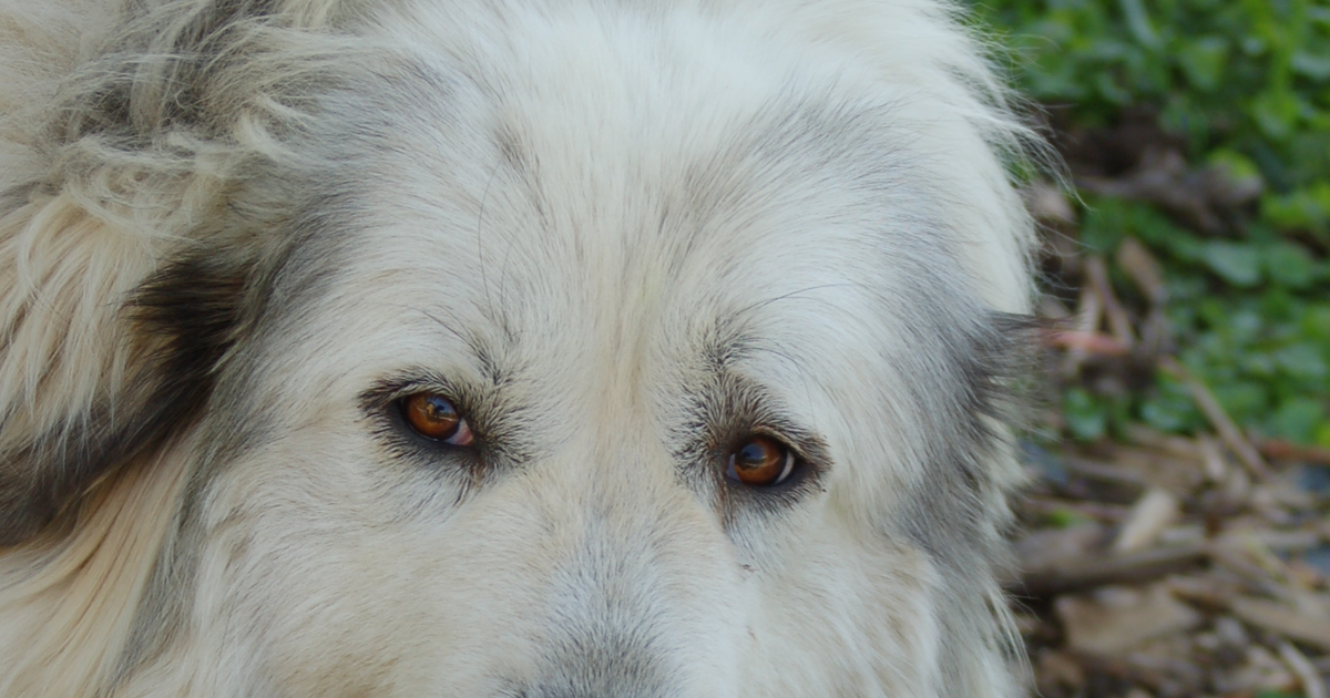
Eyecatch photo: Great Pyrenees by Carly & Art, CC BY-SA 2.0, via Wikimedia Commons.
How to get your pet tested
Some pet DNA tests screen for hereditary-disease carrier status or genetic risk markers, but the results are information, not a diagnosis. If your pet has symptoms or you need a confirmed diagnosis, please consult your veterinarian.
Below is where Canine Multifocal Retinopathy (BEST1) can be tested, grouped by where you live and marked by whether each service explicitly lists this variant (✅ = listed / ❓ = unverified / ❌ = not offered).
In the United States
In the United Kingdom
In India
Elsewhere
Note: even if the kit can be purchased/shipped internationally, the service itself (sample return, analysis, results) is not guaranteed in your country. Check each service’s stated service area and sample-return method before ordering.
Services offered in other regions (may not be available where you live)
Worried about your pet’s health? — Talk to a veterinarian
A confirmed diagnosis and any treatment plan are decisions for a veterinarian, not a test kit. The links below are professional resources.
AVMA — Find a veterinarian (American Veterinary Medical Association)
This section contains advertising (affiliate links); we may earn a commission if you buy through them. As an Amazon Associate, we earn from qualifying purchases. Genetic tests do not guarantee the prevention, diagnosis, or treatment of any disease — results indicate tendencies and provide information only.
This page is educational information, not veterinary diagnosis or advice. Always consult a veterinarian about your pet’s health.
About the author
Elena Marsh
Editor & writer (not a veterinarian)
A writer with a molecular-biology background and a lifelong dog and cat owner. Not a veterinarian — she translates peer-reviewed genetics research and primary data into plain language, always as information rather than diagnosis.